

Medical Procedure Animation: How to Show What a Camera Cannot Film
Medical procedure animationhow to show what a camera cannot film
Inside the body, a sterile theatre, patient consent and no second take. Four reasons a procedure cannot be filmed — and what a 3D model does in that place.
Shots from medical, dental and clinical animations produced by Modelight.
Some products only need to be filmed well. Others stop the camera at the patient’s skin or at the plastic housing, while everything that matters happens a few millimetres further in. This piece is about the second kind: procedures and medical devices where a photograph and a film simply do not reach.
The point is not that animation looks nicer. The point is that in this particular place it is the only material that can be produced at all.
In short: a medical procedure usually cannot be filmed for four reasons at once — the camera cannot get inside the body or inside the device, a sterile theatre has no room for a crew, a patient’s likeness requires consent, and a procedure cannot be repeated for a second take. A 3D model sidesteps all four: it cuts through tissue, is produced away from the theatre, depicts no real person, and can be corrected without going back to the clinic. The camera keeps what it is better at: the specialist’s face, the device in the hand, the trade-show report.
In this article:
- Four barriers a camera will not cross
- Five layers a needle passes through
- When the camera wins — and when animation is a waste of money
- A medical device: the mechanism that fires in a fraction of a second
- What you can show once the limits are gone
- One 3D model, four different audiences
- What the camera shows, and what the model shows
- What to send so the animation matches reality
- Frequently asked questions
Four barriers a camera will not cross
When a device manufacturer or a clinic says “let’s film how it works”, it usually turns out a few days later that there is nothing to film. There are four barriers, and they rarely come one at a time.
1. It happens on the inside
An implant driven into bone, a membrane sealing the lumen of a cannula, a compression layer working on oedema — all of it happens under tissue or under a housing. The camera reaches the surface of the material and stops. Even an endoscope gives one viewpoint at one magnification, with no way to freeze mid-movement and peel back the layer below.
2. A sterile theatre cannot be turned into a film set
Sterility comes first, and next to a patient what matters is the procedure, not the lighting. A camera operator, a tripod, extra lamps and a request for one more take are things nobody will agree to in that room — rightly so. A film set needs exactly what an operating theatre cannot have.
3. A patient is not an actor
Filming a real procedure means a specific person’s likeness, consent forms, documentation and limits on where the footage may later be shown. A 3D model depicts nobody. The same animation travels to a trade show, a clinic, a website and a distributor in another country without reopening the consent question.
4. There is no second take
A procedure takes as long as it takes, and you do not repeat it because the focus drifted. In an animation the critical moment can be slowed down, frozen, rotated and shown again from the other side — and if one component changes six months later, you fix the scene instead of organising a new shoot.
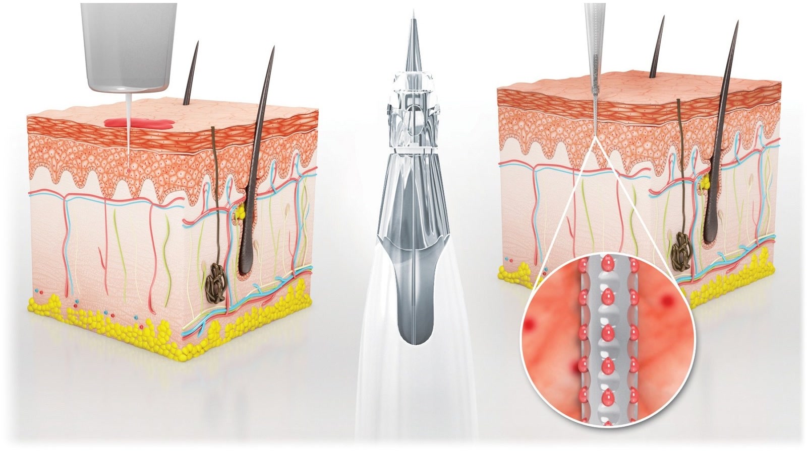
A fragment of an instructional animation for a Sol-Millennium device. The layer labels are part of the original material.
Five layers a needle passes through
This single shot explains why manufacturers of single-use devices commission an animation instead of photographs. An injection given in a clinic looks the same from the outside every time — the entire difference between devices plays out a few millimetres under the skin, where no lens reaches.
The cross-section names the layers one after another: the epidermis, below it the dermis with vessels and hair follicles, then subcutaneous tissue, the fat layer, and under it muscle and the periosteum. For the manufacturer and for whoever gives the injection this is not an anatomical detail: the layer the needle tip stops in decides whether the dose behaves as intended.
The animation does three things here at once that neither a photograph nor a leaflet diagram can. It shows depth, not just the moment of the puncture. It shows the angle of entry and how the path changes when that angle changes. And it shows scale — a difference of a few millimetres is visible in a cross-section and completely invisible in footage shot in a clinic.
Material like this outlives the campaign it was made for: it ends up in staff training, in distributor packs and in sales support in new countries, because an image needs no translation. The same logic drives explainer and instructional 3D animation — with one difference: here the alternative is not a weaker film, it is no film at all.
When the camera wins — and when animation is a waste of money
A 3D studio claiming animation always wins is selling itself rather than a solution. There are situations where ordinary footage works better and costs less.
A specialist’s face and voice build trust in a way no render can fake. If the material has to convince a doctor that a device can be trusted, another doctor who uses it and says so on camera will do more than the best animation ever made. Handling the device in a clinic — unpacking, grip, ergonomics, how it sits in the hand — also belongs on video, because that is precisely about contact with a real object. The same goes for a trade-show or installation report.
The line is simple: the camera shows what the device looks like and how it is used. The 3D model shows what happens inside it and why that works. The strongest sales materials combine both — and it pays to decide up front which part of the story goes to the film set and which to animation. If the open question is the technique itself, we settled that separately in 3D or 2D explainer video.
A medical device: the mechanism that fires in a fraction of a second
A separate category is not procedures but medical devices themselves — and here the barrier is even more literal. A needle-safety mechanism fires once, in a fraction of a second, entirely inside a plastic housing. Even if it could be filmed, there would be nothing to see but the housing moving.
In the animations of Sol-Millennium devices, for which we produced material across many products, that moment is broken down step by step: what triggers the guard, which way the component travels, and exactly when the needle stops being exposed. Worth adding: this client’s products won an iF Design Award and a Red Dot Award in 2022 — the awards went to the products, and we visualised the award-winning devices.
A fragment of a Sol-Millennium device animation — the same material serves as staff training and as an argument in a sales conversation.
For a sales team this is the difference between “our device has a safety guard” and showing exactly how that guard works — the difference between a claim and evidence. From a production standpoint it is the same work as technical animation for industry; only the scale differs, because here the whole mechanism fits into a few millimetres.
Got a medical product or a procedure that cannot be filmed?
Send a CAD file or a description of the steps — we will tell you plainly what 3D can show and what will work better on video. A conversation, not a brief.
Tell us about your productbiuro@modelight.pl | +48 509 510 197
What you can show once the limits are gone
A 3D model is not built from light bouncing off a surface — it is built from geometry, and that is the whole difference. Since the model knows where one layer ends and the next begins, that layer can simply be switched off or made transparent.
In the project for Almadent, a company in dental implantology, the procedure was shown exactly that way: transparent tissue, cross-sections opening layer by layer, and a clean environment instead of footage from a treatment room. The material explains the procedure to the specialist and to the patient, and there is not a drop of blood in it. This is not prettifying reality — there is simply nothing to hide, because none of that tissue is real.
The same technique works outside medicine, and that is where our specialisation comes from. A cross-section through a heat exchanger, through a window profile or through a mattress is exactly the same job on a different material — several dozen shots like that are collected in our cross-section gallery.
A dental implant in cross-section — an animation for Apollo Implant Components. A camera would stop at the gum; here the bone layer is opened up in motion.
One 3D model, four different audiences
A medical animation is rarely produced for a single channel. The same 3D model usually serves four completely different conversations — and that is what decides whether the project pays for itself.
Who the same material speaks to
The scenes are built once. What changes is pace, length and the descriptive layer — depending on who is on the other side of the screen.
- The specialistA doctor, a technician, a dental lab. They care about the order of steps, tolerances and how the device differs from the one they use today. Slower pace, technical labels, the full cross-section.
- Sales and distributionThey need a short version that holds up on a tablet in a clinic and needs no interpreter. The same material, trimmed and without captions, travels to any market.
- The patientA version without jargon and without views that cause anxiety. Animation has an advantage here for a simple reason: it shows the course of the procedure without showing the procedure.
- Training and trade showsThe same set of scenes works as training material for representatives and as a loop on a stand screen. Nothing has to be reshot.
Almost every medical project we deliver ends up in at least two of these uses — and that is the point where it starts paying back. A broad overview of such work is in medical and beauty projects.
What the camera shows, and what the model shows
| Situation | Camera | 3D model |
|---|---|---|
| Inside a body or a device | never gets past the surface | cross-section and transparency at any moment |
| Sterile treatment room | needs consent, a crew and concessions on sterility | produced away from the room, no effect on the procedure |
| A patient’s likeness | consent, documentation, distribution limits | depicts no real person |
| A change to the device | a new shoot | a corrected scene in the same project |
| Ergonomics and grip | shows it best | looks artificial |
| Trust in the brand | a specialist’s face and voice | will not build a relationship on its own |
What to send so the animation matches reality
The biggest risk in a medical project is not visual but factual: a beautiful scene showing a step in the wrong order is worse than no material at all. That is why we always start from four things.
A CAD model of the device — if one exists, the animation is built on real geometry rather than an approximation, and the work usually goes faster. The procedure written out step by step, ideally by the person who performs it. The audience — material for a doctor looks different from material for a patient. Regulatory limits — what may be shown and which claims must not be made; the cheapest way to avoid rework at the end.
The rest is on us: the script, the choice of shots and the decision about what to open in cross-section and what to leave in a general view. A realistic budget you can work out on our pricing page, calculator included.
See how this looks on finished projects
Implantology, single-use devices, compression dressings, tissue cross-sections — a full overview of our work for the medical and beauty sector.
Medical portfoliobiuro@modelight.pl | +48 509 510 197
Frequently asked questions
How does a medical procedure animation differ from an ordinary instructional film?
An instructional film shows actions that can be performed in front of a camera. A procedure animation reaches where no camera can be placed — under tissue, inside a housing, into a moment lasting a fraction of a second. In practice both are often made, and the dividing question is whether the important part happens on the surface or inside.
Can this kind of material be shown to patients?
Yes, and it is one of the more common uses. The animation shows the course of a procedure without showing a real procedure — no blood, no patient and none of the views that cause anxiety. Usually two versions are prepared: a full one for the specialist and a simplified one, free of jargon, for the patient.
Is a CAD file of the device required?
Not required, but it helps a great deal. With CAD we work on real geometry, so proportions, mechanism and tolerances match the product. Without it we rebuild the model from drawings, photographs and documentation, which is workable but needs more rounds of approval.
Will animation replace footage shot in a clinic?
It will not, and it should not. Video is better at ergonomics, at the device in the hand and at a specialist speaking on camera, which is what builds trust. Animation takes over what the camera cannot show. The most effective sales materials combine both sources in one edit.
Is one project enough for several markets and languages?
Yes — it is one of the main advantages of the format. Scenes are produced once, while captions, voice-over and edit length are interchangeable. The same animation serves the website, the trade-show stand, the sales presentation and training, without a second production.